Indications
- Recurrent monomorphic VT with structural heart disease, particularly with ICD shocks
- VT storm — three or more sustained episodes in 24 hours despite optimal medical therapy
- Drug-refractory or drug-intolerant VT
- Idiopathic VT (RVOT, LVOT, fascicular) — often first-line given high success and low risk
- Bundle branch reentry VT — curable with right bundle ablation
- Incessant VT causing tachycardia-mediated cardiomyopathy
Pre-procedure prep
- Imaging: TTE, cardiac MRI with gadolinium for scar characterization (especially in non-ischemic CM), coronary anatomy review
- ICD interrogation: review stored EGMs for VT morphology and cycle length; program to monitor-only or extend detection during ablation
- Antiarrhythmics: amiodarone often continued; mexiletine may be added to facilitate VT slowing for mapping
- Anticoagulation: hold DOAC per usual protocol; continuous heparin during LA or LV work
- Anesthesia: general anesthesia, arterial line, large-bore venous access, defibrillator pads
- Hemodynamic support planning: discuss Impella or ECMO availability for high-risk cases (low EF, history of unstable VT)
- Multidisciplinary discussion for complex cases — heart failure, CT surgery backup, anesthesia
Setup & equipment
- Vascular access: bilateral femoral venous, femoral arterial for LV mapping
- ICE for chamber visualization, papillary muscle and septal anatomy, effusion surveillance
- Heparin to ACT 300–350 for LV work
- Multipolar mapping catheter essential for high-density substrate maps
Technique
Mapping strategy by substrate
Idiopathic outflow tract VT
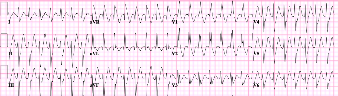
- Induce VT with isoproterenol, programmed stimulation, or burst pacing
- Activation mapping during tachycardia — find the earliest site (often 20–40 ms pre-QRS)
- Pace-map the target site and confirm 12/12 lead match with clinical VT
- Ablate at the site of earliest activation
- Common locations: RVOT septum, LVOT (aortic cusps, LV summit), pulmonary artery, papillary muscles
Scar-mediated VT (ischemic CM)
- Build a bipolar voltage map in sinus rhythm: <0.5 mV is dense scar, 0.5–1.5 mV is border zone
- Identify late potentials and local abnormal ventricular activities (LAVA) within the scar
- Channel identification: bands of preserved voltage within scar that house the reentry circuit
- Substrate modification: ablate all late potentials and channels, with or without inducing VT
- Increasingly the preferred strategy because it avoids dependence on tolerated VT mapping
Non-ischemic cardiomyopathy
- Substrate often midmyocardial or epicardial — integrate MRI scar imaging into the map
- Endocardial-only ablation often fails; epicardial access frequently required
- Common patterns: basal lateral (Chagas-like), septal (sarcoid), free wall
Bundle branch reentry
- His and right bundle recording essential
- Diagnosis: VA dissociation absent, HV during VT longer than sinus
- Ablate the right bundle — small lesion, definitive cure
- Counsel about increased likelihood of needing CRT given underlying LV dysfunction
Activation and entrainment mapping (when VT tolerated)
- Identify mid-diastolic potentials
- Entrain from candidate sites: PPI – TCL <30 ms identifies the circuit
- Concealed entrainment with matching stim-to-QRS interval identifies the critical isthmus
- Ablate during VT; termination during energy delivery within 10–20 s is highly predictive of success
Epicardial access
- Sosa subxiphoid approach under fluoroscopy
- Tuohy needle, micropuncture wire, dilator, then sheath
- Always confirm pericardial entry with contrast before sheath placement
- Map coronaries and phrenic nerve before ablation — pace-map for phrenic capture and inject contrast for coronary proximity
- Lower power settings on the epicardium; cool tip irrigation
Endpoints
- Non-inducibility of any sustained VT with aggressive programmed stimulation (up to triple extrastimuli from two sites at two cycle lengths, with isoproterenol)
- Elimination of all late potentials within the substrate
- Pace-mapping mismatch at targeted sites
- Acknowledge that complete non-inducibility isn’t always achievable — partial substrate modification still reduces ICD shock burden
Complications
- Vascular complications: ~2–3%, higher with multiple accesses and arterial work
- Tamponade: ~1–2%, higher with epicardial cases
- Stroke: 1–2% in LV mapping cases
- Heart block: variable, depends on substrate location
- Acute hemodynamic decompensation: real risk in unstable substrate — plan support upfront
- Phrenic nerve injury and coronary injury in epicardial cases
- Mortality: 1–3% in published series, concentrated in VT storm and advanced HF populations
Post-procedure care
- ICU or step-down monitoring for the first night, longer for high-acuity cases
- Telemetry for at least 24 hours; reprogram ICD before discharge with detection zones restored
- Anticoagulation for 4–6 weeks after LV ablation
- Antiarrhythmics: often continued, with reassessment at 6–12 weeks
- Heart failure optimization continues — VT ablation is not a substitute for guideline-directed therapy
- Follow-up in 2–4 weeks with device interrogation, then 3 months, then per ICD shock surveillance
- Counsel on realistic expectations: substantial reduction in shocks is the typical outcome; complete VT freedom is less common in advanced substrate