Mechanism
Atypical flutter is a macro-reentrant atrial tachycardia using a circuit other than the CTI. Most live in the left atrium and depend on prior ablation lesions, scar, or anatomic obstacles.
Common circuits
- Peri-mitral flutter: rotates around the mitral annulus. Cycle length usually 250–300 ms. Often emerges after roof or anterior LA ablation. Mitral isthmus line required to terminate.
- LA roof-dependent: travels around one or both pulmonary vein pairs through the roof. Roof line is the target.
- Anterior LA / scar-mediated: uses a region of fibrosis on the anterior wall. Increasingly recognized with mapping.
- Septal flutter: complex circuits involving the septum, sometimes incorporating the right atrium across a Bachmann’s bundle connection.
- Right atrial atypical: peri-tricuspid above the CTI, around scar from prior ASD repair, around the SVC, or upper loop reentry through the crista.
Settings to expect atypical flutter
- After AF ablation, especially with linear lesions
- After surgical maze procedures, Cox-maze IV, mini-maze
- After cardiac surgery for valve repair, CABG, ASD/VSD closure
- Congenital heart disease (Mustard, Senning, Fontan)
- Significant atrial scarring from longstanding AF without prior ablation
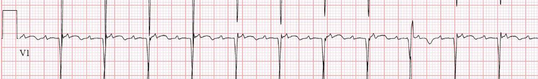
ECG features
The 12-lead is less reliable here than in typical flutter, but a few patterns help.
- Positive flutter waves in inferior leads suggest a non-CTI circuit
- Peri-mitral: positive in V1 throughout precordium, low-amplitude or notched waves in lead I
- LA roof-dependent: variable, often low-amplitude inferior waves with positive V1
- Right atrial atypical: can mimic typical but morphology differs subtly — always confirm at EP study
P-wave morphology can shift mid-tachycardia if the circuit reorganizes — common in long-standing persistent AF substrate.
Lab setup
This is where atypical flutter diverges sharply from CTI flutter.
- Transseptal access: nearly universal. Single or double puncture depending on operator preference and number of catheters needed.
- High-density mapping catheter: PentaRay, HD Grid, Octaray, or similar. Multipolar splines collect thousands of points and reveal the full circuit.
- 3D mapping system: required. CARTO or EnSite Precision. Voltage maps identify scar; activation maps trace the circuit.
- Decapolar in CS: still useful as reference and to assess LA activation
- Irrigated ablation catheter with contact force sensing
- Esophageal temperature probe for posterior LA / mitral isthmus work
- ICE catheter helpful for transseptal and ongoing monitoring
Expect a 3–5 hour case, sometimes longer.
Mapping strategy
Atypical flutter mapping is a discipline of its own.
- Voltage map in sinus or paced rhythm if possible — identifies scar and likely circuit boundaries
- Activation map during tachycardia covering ≥90% of the atrial cycle length — anything less suggests the circuit isn’t fully captured
- Entrainment from multiple sites to confirm the circuit:
- Post-pacing interval (PPI) within 30 ms of TCL → in the circuit
- PPI >30 ms → outside the circuit (bystander)
- Concealed fusion confirms entrainment from a critical isthmus
- Look for a protected isthmus — a narrow channel of slow conduction between scars or anatomic obstacles. This is the ablation target.
- Differentiate macro-reentry from focal with localized vs distributed activation
Ablation
- Peri-mitral: mitral isthmus line from the left inferior PV to the mitral annulus. Often needs CS ablation from inside the vein to achieve block.
- Roof-dependent: linear lesion across the LA roof connecting the superior PVs.
- Scar-mediated: target the protected isthmus identified by mapping.
- Endpoint: termination during ablation plus demonstrable block across the ablation line, plus non-inducibility on burst pacing.
Why outcomes are humbler
- Multiple potential circuits in the same substrate
- Reaching complete block across long linear lesions is technically demanding
- Mitral isthmus block requires CS ablation in ~50% of cases
- New circuits can emerge from ablation lines themselves
- Acute success 70–85%; 12-month freedom from atrial arrhythmia 50–70%
Practical notes
- Counsel realistically — this is not a one-and-done procedure for most patients
- Anticoagulation: as for AF, continued indefinitely in most
- Antiarrhythmics often continued post-ablation, at least short-term
- Document the circuit and ablation lines meticulously — the next operator will need it