Focal AT
A discrete site fires repetitively at a rate higher than sinus. Three mechanisms operate, often indistinguishable on the surface ECG.
- Automaticity: enhanced spontaneous depolarization. Warm-up at onset, cool-down at termination, sensitive to catecholamines. Often comes and goes — frustrating to capture in the lab.
- Triggered activity: from delayed afterdepolarizations. Adenosine-sensitive in many cases. Can mimic the others.
- Micro-reentry: small reentrant circuit, can be entrained. More likely after atrial surgery, ablation, or scar.
Common sites
- Right atrium: crista terminalis (most common), tricuspid annulus, CS os, right atrial appendage, perinodal area
- Left atrium: pulmonary vein ostia (especially post-AF ablation), mitral annulus, left atrial appendage, septum, LA roof
- The septum and perinodal region are tricky — ablation risk to the AV node is real
ECG localization
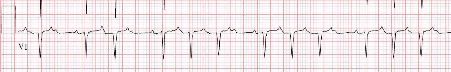
- Lead V1: positive P → left atrial; negative or biphasic → right atrial
- Lead aVL: negative P → left atrial; positive → right atrial
- Inferior leads: positive → superior focus (crista, RUPV); negative → inferior focus (CS os, low crista)
- Lead I: negative → left lateral; positive → right or septal
P morphology is best assessed during AV block (adenosine, beta-blocker bolus) or in patient’s own tachycardia at slow conduction.
Multifocal atrial tachycardia (MAT)
Not a reentrant arrhythmia and not an ablation target. MAT reflects atrial sickness from a systemic insult.
- Setting: severe COPD exacerbation, hypoxia, theophylline or beta-agonist toxicity, sepsis, hypomagnesemia, post-cardiac surgery, decompensated heart failure
- ECG criteria: ≥3 P-wave morphologies in the same lead, irregular PP and PR intervals, isoelectric baseline (unlike AF)
- Treatment:
- Fix the underlying illness — oxygenation, magnesium repletion, treat infection, dial back bronchodilators
- Magnesium IV often helps even with normal serum levels
- Metoprolol IV if tolerated (caution in bronchospastic disease) or diltiazem
- Amiodarone occasionally; cardioversion is futile (rhythm is automaticity-based)
- No role for ablation
Distinguishing AT from atypical flutter
This trips people up regularly.
- AT: discrete P waves with an isoelectric baseline between them
- Atypical flutter: continuous undulating atrial activity, no flat baseline
- AT cycle length is usually 250–500 ms; flutter typically 200–300 ms but overlaps
- Atypical flutter is often post-ablation or post-surgical; AT can occur in normal hearts
- On the table, both behave differently — flutter is a macro-reentrant circuit you can entrain around an anatomic obstacle; focal AT entrains from one site only or doesn’t entrain at all
EP study and ablation
- Activation mapping: identify the earliest activation site relative to the P wave onset. Look for “pre-P” activation — typically 20–40 ms before P onset at the source.
- 3D mapping system (CARTO or EnSite) is standard. High-density mapping pays off in low-amplitude or intermittent ATs.
- Pace mapping at the suspected focus reproduces the P-wave morphology in focal AT.
- Entrainment is useful for micro-reentry but not for true focal mechanisms.
- Ablation: focal lesion at the earliest site. Endpoint is non-inducibility on full isoproterenol challenge.
Practical pitfalls
- Catecholamines required to induce — have isoproterenol ready
- Burst pacing and PES can initiate or terminate
- Septal and para-Hisian sites: cryo, or RF with constant monitoring of PR/AH
- Always think about the PVs in anyone with prior AF ablation — a “focal AT” off the LSPV is really a reconnected PV