Mechanism
AF is the end result of two interacting forces: triggers that initiate the arrhythmia and substrate that sustains it.
- Triggers: ectopic firing from myocardial sleeves extending into the pulmonary veins accounts for the majority of paroxysmal initiations. Non-PV triggers — superior vena cava, coronary sinus, ligament of Marshall, left atrial appendage, crista terminalis — become more relevant in persistent disease and after a failed first ablation.
- Substrate: atrial stretch, fibrosis, inflammation, and autonomic remodeling shorten refractoriness and create regions of slow, anisotropic conduction. Rotors and multiple wavelets propagate through this tissue.
- Remodeling is bidirectional: AF begets AF. Each episode shortens atrial refractoriness within hours and promotes fibrosis over months. This is why early intervention matters.
Classification by burden
- Paroxysmal: self-terminates within 7 days (usually within 24 h)
- Persistent: sustained beyond 7 days or requires cardioversion
- Long-standing persistent: continuous AF more than 12 months
- Permanent: a shared decision to abandon rhythm control, not a mechanistic category
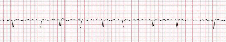
ECG features
The hallmark is irregular irregularity without organized atrial activity. Look at lead V1 for the cleanest view of fibrillatory waves; coarse f-waves can mimic flutter but lack the consistent sawtooth and cycle length. Watch for hidden flutter in patients on antiarrhythmics — class IC drugs in particular can organize AF into a slow, 1:1-conducting atypical flutter.
A regularized AF rhythm in a previously irregular patient should prompt one of three thoughts:
- Complete heart block with junctional or ventricular escape
- Conversion to atrial tachycardia or flutter
- Ventricular paced rhythm
Work-up
- 12-lead ECG to confirm and exclude pre-excitation
- TTE for chamber sizes, LV function, valvular disease, pericardial effusion
- Labs: TSH, CBC, BMP, magnesium
- Ambulatory monitoring when paroxysmal and capture is needed: patch monitor for symptom correlation, longer monitors or implantable loop recorders for cryptogenic stroke work-up
- Sleep evaluation — OSA is massively under-diagnosed in our AF population and a top driver of recurrence
- TEE or cardiac CT for pre-ablation planning and LAA thrombus exclusion
Treatment overview
- Rate control: beta-blockers, non-DHP calcium channel blockers, digoxin in selected patients. RACE II target is a resting rate under 110 if asymptomatic.
- Rhythm control: cardioversion, antiarrhythmics (flecainide and propafenone in structurally normal hearts; sotalol, dofetilide, amiodarone otherwise), and catheter ablation.
- Anticoagulation: DOAC preferred over warfarin in non-valvular AF. CHA2DS2-VASc ≥2 in men or ≥3 in women drives treatment. LAA occlusion for patients with absolute or relative contraindications to long-term anticoagulation.
- Risk factor modification: weight loss, OSA treatment, BP control, alcohol reduction, exercise. The LEGACY data are persuasive — patients who lose >10% body weight have markedly better ablation outcomes.
What we do in clinic
For a new AF referral, the first visit covers:
- Confirming the arrhythmia and characterizing the burden
- Stroke risk stratification and anticoagulation decision
- Symptom assessment — many “asymptomatic” patients describe substantial fatigue once asked specifically
- A frank conversation about early rhythm control. We favor ablation early in symptomatic paroxysmal AF; EAST-AFNET 4 and CABANA subgroup data support this.
- Setting up a sleep study if there’s any clinical suspicion
Repeat ablations are common (15–25% at 1–2 years) and usually reveal PV reconnection or new non-PV triggers. We counsel patients up front so a touch-up isn’t framed as failure.